
What is Women’s Imaging?
Radiology Associates of New Hartford partners with area medical providers to care for women’s healthcare needs in the Mohawk Valley. Women’s Imaging encompasses obstetrical, gynecological, bone density, and breast imaging. Our fellowship-trained radiologists specialize in leading-edge breast imaging procedures, including digital mammography, breast ultrasound, and breast MRI. Our physicians also perform breast biopsies and interpret bone densitometry (DEXA) exams.
Mammography is used as both a screening and a diagnostic tool. Screening serves as a routine check-up for at-risk patients that are not currently experiencing any symptoms. Diagnostic mammograms allow us to investigate lumps or other potential signs of breast cancer using low-dose x-ray to visualize breast tissue. This method reveals tissue abnormalities such as cysts or cancerous growths. Mammography, combined with a woman’s normal self-breast exams, remains the most effective screening tool for detecting breast cancer early.
Breast ultrasound uses sound waves to obtain images of breast tissue using a handheld device called a transducer. This method allows your technologist to identify whether an area of interest is a fluid-filled cyst or a solid mass (tumor) as the sound waves reflect an image back to the workstation.
Following a mammogram and/or ultrasound, your radiology team may recommend a biopsy of any areas of concern in your breast. This small tissue sample, observed under a microscope, allows your doctor to reliably determine if your abnormalities are benign or cancerous. Typically these are non-surgical, outpatient procedures, simply using a localized anesthetic and a needle to aspirate the tissue as long as the abnormality is a palpable lump. Biopsies cause little to no pain, though slight subsequent bruising and soreness is not uncommon. Depending on the location, size and characteristics of your abnormality, your doctor may recommend surgical excisional biopsy with needle localization, or prefer to use an MRI, ultrasound or mammogram to assist in visualizing the area. At Radiology Associates of New Hartford, we choose stereotactic breast biopsy whenever surgery is not required, as it ensures the best results and experience for our patients. This procedure uses a number of computerized mammogram images to identify a breast abnormality in three dimensions for biopsy.
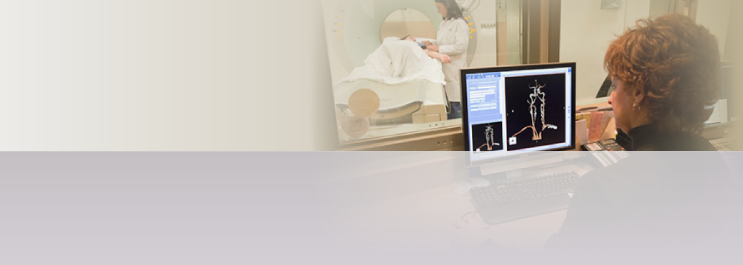
Breast MRI (Magnetic Resonance Imaging), like all MRI procedures, uses a magnetic field and radio waves to develop a full, cross-sectional picture of the breasts. As it uses no x-rays or radiation, MRI has no known side effects, making it one of the safest, most advanced diagnostic imaging tools available to doctors today. Breast MRI complements mammography and stereotactic breast biopsy and may be used to locate, define and/or confirm cancerous metastasis as well as implant leakage or rupture. It does not replace routine mammography, breast self-examination (BSE), physician check-ups, or biopsy. You may not be eligible for breast MRI if you cannot lie on your stomach for 30 minutes or more, have metal implants or fragments of any kind, including pacemakers, or are pregnant.
Bone Densitometry exams, also known as DEXA scans (dual energy X-ray absorption), remain the safest, most-effective tool for diagnosing osteoporosis. This painless procedure measures bone density to determine a patient’s risk of osteoporosis.